Intratesticular Epidermoid Cyst Masquerading as Testicular Torsion
Jeremy Slawin, BA,1 Kevin Slawin, MD2
1 Baylor College of Medicine, Texas Medical Center, Houston, TX and Rice University, Houston, TX; 2Vanguard Urologic Institute at Memorial Hermann Medical Group, Houston, TX, Baylor College of Medicine, Texas Medical Center, Houston, TX, and Memorial Hermann Hospital, Texas Medical Center, Houston, TX
Epidermoid cysts are benign tumors that comprise approximately 1% of all testicular masses. They usually present as painless masses that can be identified on scrotal ultrasound as well-demarcated intratesticular lesions with mixed echogenicity. This case report describes a rare presentation of an extremely large intratesticular epidermoid cyst with clinical and radiologic findings more consistent with testicular torsion. The sizeable cyst obliterated the surrounding testicular parenchyma, causing it to appear on scrotal Doppler ultrasound as a testicle devoid of blood flow. This obliteration also resulted in failure to identify a testicular mass on physical examination or imaging. The current literature contains previous reports of extratesticular epidermoid cysts presenting as torsion; however, this is the first report of an intratesticular epidermoid cyst presenting in this manner. Though smaller cysts may be managed effectively with testicular-sparing surgery, optimal management of a cyst this size requires orchiectomy.
[Rev Urol. 2014;16(4):198-201 doi: 10.3909/riu0633]
© 2014 MedReviews®, LLC
Key words
Epidermoid cyst • Testicular torsion • Acute testicular pain • Intratesticular • Doppler ultrasound
Key words
Epidermoid cyst • Testicular torsion • Acute testicular pain • Intratesticular • Doppler ultrasound

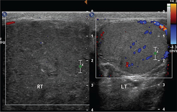
Figure 1. Doppler ultrasound revealed an enlarged right testicle devoid of blood flow. The left testicle appeared normal.

Figure 1. Doppler ultrasound revealed an enlarged right testicle devoid of blood flow. The left testicle appeared normal.

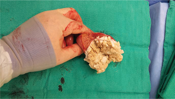
Figure 2. The right testicle was nearly entirely comprised of caseous, keratinous debris, the contents of a typical epidermoid cyst.

Figure 2. The right testicle was nearly entirely comprised of caseous, keratinous debris, the contents of a typical epidermoid cyst.
In this case of an intratesticular epidermoid cyst presenting as testicular torsion, this important clinical clue—an extra testicular mass—was absent, making it a unique diagnostic dilemma…
… because the entire testicle was comprised of the cyst, no distinct mass could be appreciated on examination or ultrasound.
Main Points
• A large intratesticular epidermoid cyst can mimic the clinical presentation of testicular torsion. If sufficiently large, it may compress and obliterate the surrounding testicular parenchyma, resulting in pain, the absence of a mass on physical examination, and an appearance on scrotal Doppler ultrasound of a testicle devoid of blood flow.
• Intratesticular epidermoid cysts usually present as small, painless testicular masses that are discovered on a routine self- or physical examination. Systemic symptoms are typically absent. In some cases, they can be treated with testicle-sparing surgery such as tumor enucleation and wedge excision, although these approaches are unlikely to be successful in a large cyst. Furthermore, due to symptomatology that mimics malignant pathology, radical inguinal orchiectomy is often performed instead.
• Inclusion of intratesticular epidermoid cysts on the differential of suspected torsion is worthwhile so they can be identified and managed appropriately. Smaller cysts may be effectively managed with testicle-sparing surgical techniques, whereas very large cysts may require orchiectomy.
Intratesticular epidermoid cysts are relatively rare benign testicular masses that comprise approximately 1% of all testicular tumors.1-6 An epidermoid cyst typically presents as a painless testicular mass and consequently often mimics the presentation of a malignant testicular neoplasm. This case review details the case of a patient with an unusual presentation of an intratesticular epidermoid cyst—one of acute testicular torsion. To our knowledge, this is the first case of its kind to be reported in the literature.
Case Report
A 61-year-old man presented to the emergency department complaining of an acute onset of right testicular pain. A scrotal Doppler ultrasound was performed before the urologic team arrived to evaluate the patient. The ultrasound revealed an enlarged right testicle measuring 5.9 cm × 4.8 cm × 5.1 cm that was devoid of blood flow (Figure 1). In addition, the radiologist noted that no discrete masses were present. Accordingly, the radiologist made a preoperative diagnosis of right testicular torsion. The urology team subsequently performed a physical examination, which revealed an exquisitely tender and swollen right testicle with normal lie. No masses were palpated. These findings were consistent with the diagnosis of testicular torsion, and the patient was taken immediately to surgery.
A midline scrotal incision was made to enter the scrotum. The right testicle was delivered through this incision, and the right cord was serially dissected until the right testicle was freely mobile. The right testicle appeared dusky and necrotic, and a right orchiectomy was subsequently performed to remove what seemed to be an unsalvageable right testicle. Off the table, the testicle was opened, revealing a large, caseous, necrotic-appearing mass within the testicle (Figure 2).
The specimen was sent for pathologic analysis. The mass was determined to be a well-circumscribed epidermoid cyst, measuring 6.6 cm × 6.0 cm × 4.0 cm with normal testicular tissue compressed at the periphery.
Discussion
An epidermoid cyst is the most common form of cutaneous cyst and usually presents as a discrete nodule on the skin of the scalp, face, neck, trunk, and back.7 Rarely, they appear as intratesticular masses or intrascrotal extratesticular masses, in total accounting for approximately 1% of all testicular tumors.1-6 Testicular epidermoid cysts clinically present as small, painless testicular masses that are typically discovered during a routine self- or physical examination. However, approximately 15% of these cysts will present with pain.5 On ultrasound, the cysts classically appear as well-demarcated masses that are hypoechoic with scattered echogenic foci or have an “onion skin” pattern with alternating hyperechoic and hypoechoic rings.3,7 Some epidermoid cysts have also been described as having similar echotexture to that of a normal testicle.2
Although our patient had an intratesticular epidermoid cyst, he lacked the classic signs and symptoms described above. Instead, his clinical presentation was more consistent with testicular torsion, because he had the characteristic symptom of torsion: an acute onset of testicular pain.8 In addition, scrotal ultrasound showed what appeared to be a large testicle devoid of blood flow. Doppler ultrasound revealing absent or minimal flow to the testicle suggests the diagnosis of testicular torsion with high sensitivity and high specificity.9 Furthermore, neither physical examination nor scrotal ultrasound was able to appreciate a testicular mass, a finding expected to accompany a testicular epidermoid cyst.5
Our patient's atypical signs and symptoms can likely be attributed to the large size of his cyst: 6.6 cm compared with the average size of 2 cm. The cyst grew so large that it occupied the majority of the testicle, likely stretching the testicular tunica albuginea and causing pain. In addition, because the entire testicle was comprised of the cyst, no distinct mass could be appreciated on examination or ultrasound. Because the cyst obliterated the surrounding testicular parenchyma, Doppler ultrasound visualized only the cyst, producing an image that had a similar appearance to a testicle lacking blood flow.
To our knowledge, this is the first reported case of an intratesticular epidermoid cyst with presenting signs and symptoms of acute testicular torsion. There are, however, two case reports in the literature that describe an extratesticular epidermoid cyst presenting as testicular torsion.10,11 An extratesticular epidermoid cyst typically forms a separate mass within the scrotum; thus, in both of the reported cases, the cysts were misdiagnosed as supernumerary testicles that had subsequently torsed. In these cases, the presence of the third mass may have led one to the correct diagnosis. Given the extremely rare nature of true polyorchidism,12 it is reasonable to consider alternate etiologies for a third testicular mass presenting with acute pain, including an extratesticular epidermoid cyst. Moreover, in true cases of triorchidism, all three scrotal masses will have been present since birth. If the third mass developed during adulthood, acquired pathology such as an extratesticular epidermoid cyst should be considered as the etiology.
In this case of an intratesticular epidermoid cyst presenting as testicular torsion, this important clinical clue—an extra testicular mass—was absent, making it a unique diagnostic dilemma, unlike other case reports. This made it even more challenging to diagnose the condition correctly preoperatively.
It is important to note that despite the incorrect preoperative diagnosis, this patient received the most appropriate treatment. The consequences of untreated testicular torsion are severe, and rapid surgical exploration is warranted even in cases that are equivocal.8 Because this patient's epidermoid cyst was so large, surgical removal would have been necessary to relieve his symptoms. Tumor enucleation and wedge resection with intraoperative frozen section have been utilized successfully to resect epidermoid cysts5,6; however, our patient's cyst obliterated nearly all of the testicular parenchyma and these approaches likely would have been unsuccessful.
Conversely, these testicle-sparing surgical techniques may be appropriate for the management of smaller intratesticular epidermoid cysts. In addition, because smaller cysts will not fully obliterate the testicular parenchyma, they will be more likely to be identified correctly on Doppler ultrasound, allowing for the development of a proper surgical plan. However, although the presence of the classic echotexture on scrotal ultrasound may suggest the diagnosis of the benign intratesticular epidermoid cyst, ultrasound cannot definitively discern between benign and malignant masses.6 This circumstance, in addition to the fact that epidermoid cysts often mimic the clinical history of malignant masses, has resulted in inguinal radical orchiectomy being the primary mode of therapy.5
It is important to include intratesticular epidermoid cysts in the differential diagnosis in suspected testicular torsion, as knowledge of this unusual presentation can aid in earlier recognition and potentially alter management. Orchiectomy can be avoided for small cysts, but orchiectomy is the proper surgical approach for large cysts that obliterate the surrounding testicular parenchyma.
Conclusions
Patients with testicular torsion typically present with acute testicular pain; scrotal Doppler ultrasound often reveals a testicle without blood flow. Rarely, as was reported in this case, a sufficiently large epidermoid cyst can be the etiology of these findings. This can occur when the cyst completely obliterates its surrounding testicular parenchyma. If identified preoperatively, small epidermoid cysts can be managed effectively with testicle-sparing surgery; however, definitive management of a very large, painful epidermoid cyst is orchiectomy. ![]()
The authors report no real or apparent conflicts of interest.
References
- Agarwal A, Agarwal K. Intrascrotal extratesticular epidermoid cyst. Br J Radiol. 2011;84:121-122.
- Kao HW, Wu CJ, Cheng MF, et al. Extratesticular epidermoid cyst mimicking enlarged testis. Ir J Med Sci. 2011;180:593-595.
- Langer JE, Ramchandani P, Siegelman ES, Banner MR Epidermoid cysts of the testicle: sonographic and MR imaging features. AJR Am J Roentgenol. 1999;173:1295-1299.
- Price EB Jr. Epidermoid cysts of the testis: a clinical and pathologic analysis of 69 cases from the testicular tumor registry. J Urol. 1969;102:708-713.
- Shah KH, Maxted WC, Chun B. Epidermoid cysts of the testis: a report of three cases and a review of the literature. Cancer. 1981;47:577-582.
- Heidenreich A, Engelmann UH, Vietsch HV, Derschum W. Organ preserving surgery in testicular epidermoid cysts. J Urol. 1995;153:1147-1150.
- Lee HS, Joo KB, Song HT, et al. Relationship between sonographic and pathologic findings in epidermal inclusion cysts. J Clin Ultrasound. 2001; 29:374-383.
- Barthold JS. Abnormalities of the testes and scrotum and their surgical management. In: Wein AJ, ed. Saunders Elsevier; 2011:3557.
- Kapasi Z, Halliday S. Best evidence topic report. Ultrasound in the diagnosis of testicular torsion. Emerg Med J. 2005;22:559-560.
- Fanning DM, McDermott T. An epidermoid cyst presenting as testicular torsion in a patient with triorchidism. BMJ Case Rep. 2011;2011:bcr0520114170.
- Graif A, Gakhal M, Iacocca MV, Levy HM. Ruptured extratesticular epidermal inclusion cyst mimicking polyorchidism with torsion on sonography [published online ahead of print May 7, 2014]. Emerg Radiol. doi: 10.1007/s10140-014-1229-x.
- Bergholz R, Wenke K. Polyorchidism: a meta-analysis. J Urol. 2009;182:2422-2427.