Ureteral Stone Associated With a Middle Blind Ending of a Bifid Ureter
Selahattin Çalişkan, MD
Department of Urology, Hitit University Çorum Training and Research Hospital, Çorum, Turkey
A blind-ending bifid ureter is an anatomic variant of ureteral duplications. There are three forms of blind-ending bifid ureter, classified depending on their location. A proximal blind-ending bifid ureter is the most common subtype of this congenital anomaly. Most patients are asymptomatic and only show symptoms after the complication has occurred. These complications include stone formation, vesicoureteric reflux, and ureteral tumor. Intravenous urography is usually used to diagnose bifid ureter; voiding cystourethrography, photofluoroscopy, cineroentgenography, cystoscopy with retrograde pyelography, computed tomography (CT), multidetector CT, and magnetic resonance urographies are other imaging methods used. This is a case of a ureteral stone associated with a middle blind ending of a bifid ureter.
[Rev Urol. 2016;18(1):54-56 doi: 10.3909/riu0688]
© 2016 MedReviews®, LLC
Key words
Blind-ending bifid ureter • Ureteral duplication • Ureteral stone • Ureteroscopic stone removal
Key words
Blind-ending bifid ureter • Ureteral duplication • Ureteral stone • Ureteroscopic stone removal
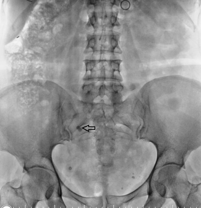
Figure 1. Ureteral stone localized in the middle ureter (arrow).
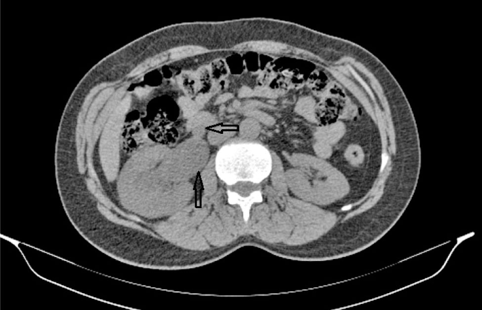
Figure 2. Arrows show the ureter and renal pelvis.
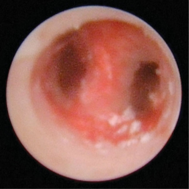
Figure 3. Endoscopic image of a bifid ureter and stone localization.
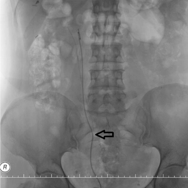
Figure 4. A double J stent and ureteral catheter were inserted in each ureter separately. The arrow shows the blind ureter location.
Recurrent urinary tract infection and poorly defined abdominal pain are the most common symptoms.
… this anomaly is often missed by radiologists because normal collecting systems are assumed if uncustomized imaging modalities are used…
Main Points
• Blind-ending bifid ureter is an anatomic variant and develops from premature division of the metanephric diverticulum, the primordium of the renal pelvis, and the ureter. The ureteral duplications are subdivided into two groups, partial or complete.
• This uncommon congenital anomaly has three subtypes—proximal, middle, and distal—depending on location; the middle blind-ending bifid ureter is the least common subtype.
• Intravenous urography is the most frequently used imaging study to diagnose a bifid ureter. Multidetector computed tomography and magnetic resonance urography are also useful diagnostic alternatives.
• Complications such as vesicoureteric reflux, stone formation, and ureteral tumor in symptomatic patients must be treated.
Ureteral duplication is one of the most common congenital anomalies of the urinary system, with an incidence of 0.8% in autopsy series.1 Herbert first described the entity of the double-blind ureter in 1904.2 There are approximately 200 reported cases in the literature.3 This rare anomaly is more prevalent in women than men and is observed almost twice as often on the right side.1 Intravenous urography is the most frequently used imaging study to diagnose a bifid ureter.3 This case report presents a patient with a middle blind ending of a bifid ureter with ureteral stone who was treated with ureteroscopic stone removal.
Case Report
A 46-year-old woman presented to our clinic with right flank pain for the previous month. She had a history of ureteroscopic stone surgery performed two times. Laboratory test results were all within normal ranges. Imaging of the kidney, ureter, and bladder showed a right distal ureter stone (Figure 1). Computed tomography (CT) revealed an 11-mm stone in the right ureter with ureteral duplication (Figure 2). Endoscopic stone removal was planned. After the stone was removed a middle blind-ending bifid ureter was confirmed (Figure 3). A ureteral catheter and double J stent were inserted to each ureter separately (Figure 4). No complications occurred during the intra- or postoperative periods. The patient was discharged 1 day after the operation.
Discussion
Blind-ending bifid ureter is an anatomic variant and develops from premature division of the metanephric diverticulum, the primordium of the renal pelvis, and the ureter.4 The ureteral duplications are subdivided into two groups, partial or complete, according to the American Academy of Pediatrics.5 Partially duplicated systems are more common than complete ones, with a ratio of 3:1.6 The blind-ending bifid ureter is three times more common in women and is diagnosed twice as often on the right side as the left.7 The bifid ureters may join and enter as a single unit to the bladder or open to the bladder with different units, separately.8 This uncommon congenital anomaly has three subtypes—proximal, middle, and distal—depending on location.1 The middle blind-ending bifid ureter is the least common subtype.
The incidence of this anomaly is low, because most patients are asymptomatic7; blind-ending ureters are usually clinically insignificant and typically do not produce symptoms. Symptomatic cases include complaints about abdominal pain, dysuria, frequent micturition, and hematuria. The complaints could be associated with stone formation, vesicoureteric reflux, or ureteral tumor.1 Recurrent urinary tract infection and poorly defined abdominal pain are the most common symptoms.3 The causes of pain are an inflammatory process in the blind-ending ureteral branch on the side of urinary retention, and peristaltic disturbances caused by ureteroureteral reflux.9
Most patients are diagnosed using intravenous urography or retrograde pyelogram.10 However, this anomaly is often missed by radiologists because normal collecting systems are assumed if uncustomized imaging modalities are used, such as intravenous urography or ultrasound.10 Other diagnostic imaging modalities include voiding cystourethrography, photofluoroscopy, cineroentgenography, cystoscopy with retrograde pyelography, or CT.3 Multidetector CT and magnetic resonance urography are also useful diagnostic alternatives.1
Complications such as vesicoureteric reflux, stone formation, and ureteral tumor in symptomatic patients must be treated.1 Vesicoureteric reflux, poor peristalsis, stasis, and recurrent infections in the blind ending of the ureter are the causes of stone formation.10 This rare entity should be considered to prevent unexpected complications during surgery.
References
- Karabacak OR, Bozkurt H, Dilli A, et al. Distal blind-ending branch of a bifid ureter. Arch Med Sci. 2013;9:188-190.
- Herbert H. Diverticule de l’uretre. Bull Med Soc Anat Paris. 1904;6:76-77.
- Chang E, Santillan C, O’Boyle MK. Blind-ending branch of a bifid ureter: multidetector CT imaging findings. Br J Radiol. 2011;84:e38-e40.
- Gandhi KR, Verma HV, Siddiqui AU, Wabale RN. A retroaortic left renal vein associated with a partially bifid left ureter-case report. Eur J Anat. 2014;18:42-44.
- Glassberg KI, Braren V, Duckett JW, et al. Suggested terminology for duplex systems, ectopic ureters and ureteroceles. J Urol. 1984;132:1153-1154.
- Bhamani A, Srivastava M. Obstructed bifid uretereric system causing unilateral hydronephrosis. Rev Urol. 2013;15:131-134.
- Kodama K, Iwasa Y, Motoi I, et al. Urothelial carcinoma associated with a blind-ending bifid ureter. International Cancer Conference Journal. 2012;1:147-150.
- Harris RD. Bifid ureter with blind-ending branch–the radiologist’s role in diagnosis. West J Med. 1975;122:320-321.
- Lenaghan D. Bifid ureters in children: an anatomical and clinical study. J Urol. 1962;87:808.
- Ustuner E, Atman ED, Yagci C, et al. US and MDCT findings in a caudal blind ending bifid ureter with calculi. Clin Pract. 2011;1:e77.